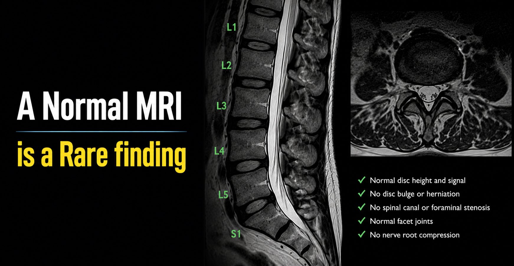
A Normal Lumbar MRI Is a Rare Finding for Anyone Older Than 40
By John H. Shim, MD, FAAOS | Orthopaedic Spine Surgeon | ShimSpine.com
If you are over the age of 40 and your lumbar MRI comes back showing degenerative changes, disc bulges, or mild arthritis — welcome to the club. You are, statistically speaking, perfectly normal. In fact, a completely “clean” lumbar MRI in a middle-aged or older adult is the anomaly, not the rule.
Degeneration Begins Earlier Than You Think
The natural history of intervertebral disc degeneration is humbling in its inevitability. Contrary to what many patients assume, the process does not begin in middle age; it begins in adolescence. By the mid-teenage years, the nucleus pulposus of the lumbar discs begins to lose hydration, and the cellular matrix that gives discs their shock-absorbing resilience starts its slow, decades-long transformation. Biochemically, the proteoglycan content decreases, collagen composition shifts, and the organized lamellar architecture of the annulus fibrosus gradually loses its pristine structure. This is not pathology. This is biology.
What the Research Tells Us
The medical literature of the past decade and a half has made one thing abundantly clear: degenerative MRI findings are the norm in asymptomatic adults, not the exception. Landmark studies published in the American Journal of Neuroradiology and The Lancet have consistently shown that disc degeneration is present in roughly 37% of asymptomatic individuals in their 20s, climbing to over 80% by age 55, and approaching near-universal prevalence after age 60. Disc bulges are found in approximately 50–60% of pain-free adults over 40. Facet arthropathy, loss of disc height, and Modic endplate changes follow similarly predictable patterns. A systematic review by Brinjikji and colleagues (2015), encompassing over 3,000 asymptomatic subjects, provided a comprehensive atlas of these age-related normative findings, and the numbers are striking in their consistency.
The MRI Is Not a Crystal Ball
Here is the analogy I often use with my patients: finding disc degeneration on a lumbar MRI after age 40 is like finding gray hair on a 50-year-old. It tells you something about the passage of time; it does not tell you why your back hurts. The MRI captures anatomy; it does not capture pain generators. Studies have repeatedly demonstrated a weak correlation between structural findings on imaging and actual clinical symptoms. A large disc bulge in one patient may cause zero discomfort, while another patient with minimal MRI changes experiences debilitating pain. The scan is a snapshot. It is not a diagnosis.
Neurological deficits, true motor weakness, loss of bowel or bladder control, and progressive myelopathy are the findings that demand urgent attention regardless of what imaging shows. Short of those red flags, the presence of degenerative change on an MRI is rarely the emergency patients fear it to be.
A More Informed Way to Think About Your Spine
Here is the bottom line, and I offer it with genuine optimism: expect your MRI to show degenerative changes. They are a known, well-documented, and largely universal expression of aging in the human spine. Unless your imaging identifies something limb-threatening or life-threatening cauda equina compression, an instability, an unexpected mass, those findings should be contextualized as part of the aging process, not as a verdict requiring immediate intervention.
The spine you carry into your 40s, 50s, 60s, and beyond is a spine that has worked hard for you. The MRI may show the miles. That does not mean you are broken.
―――
John H. Shim, MD, FAAOS is a board-certified orthopaedic spine surgeon based in Oldsmar, Florida, with over 30 years of clinical experience.
Last modified: July 28, 2026