Thoracic Spine Anatomy
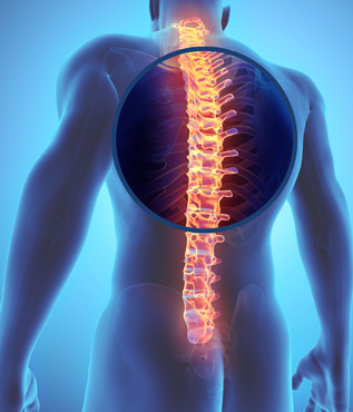 The thoracic spine is located between the cervical spine (neck) and the lumbar spine (low back). The thoracic spine is made up of 12 vertebrae, labeled T1 to T12. The ribs connect to the thoracic spine and together with the sternum (breast bone), forms the thoracic cage. This serves to protect the lungs and underlying organs.
The thoracic spine has an inward curve or kyphosis.
Similar to the rest of the spine, the thoracic spine vertebral bodies contain facet joints and feature a vertebral arch which provides space for the spinal cord. Within the thoracic spine, there are 12 pairs of spinal nerves that branch off of the spinal cord. Supporting muscles and ligaments attach to the back of the vertebral arch (spinous process) which allows for movement.
Between each of the vertebral segments lies the intervertebral disc (“disc”). The disc serves as a shock absorber and also allows for motion.
The thoracic spine is prone to a number of injuries including muscle sprains, disc herniations, and fractures (compression fractures) that result from abnormal stress placed upon weakened bones.
Last modified: October 22, 2019