Getting a Spine MRI Can Increase Your Chances of Future Surgery, Even After Your Pain Is Gone
A closer look at an underappreciated consequence of early imaging — and what it means for your long-term spine health.
What Most Patients Are Never Told About Spine MRIs
Every year, millions of patients with back or neck pain undergo MRI studies. For many, the pain resolves on its own within weeks or months. It is a natural outcome that holds true for the vast majority of acute spine complaints. The MRI, it often turns out, was not strictly necessary for recovery.
What is less commonly discussed is that obtaining a spine MRI before your pain resolved may meaningfully increase your statistical risk of undergoing spine surgery at some point in the future. Not because surgery became medically necessary, but because of a chain of clinical and psychological effects that a documented imaging finding sets in motion.
This is not an argument against MRI. MRI is an invaluable diagnostic tool for the right indications. The concern is with early or routine imaging when the clinical picture does not require it, and with the consequences that can follow once a structural finding appears in your permanent medical record.

The Problem: Normal Aging Looks Abnormal on MRI
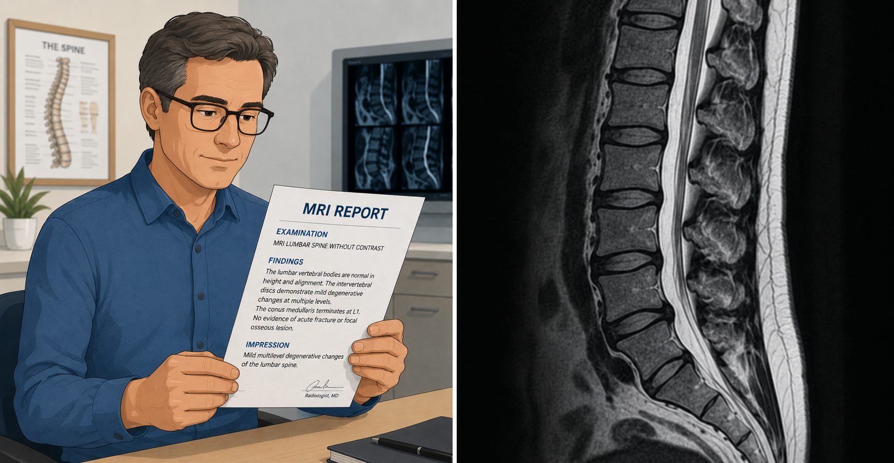
Spine MRIs are extraordinarily sensitive instruments. They detect structural changes with precision, including changes that are entirely normal for your age and have no relationship to your symptoms whatsoever.
A landmark systematic review published in the American Journal of Neuroradiology (Brinjikji et al., 2015) examined the prevalence of degenerative spine findings in asymptomatic adults. These are people with no back pain at all. The findings were striking:
- By age 40, approximately 68% of asymptomatic adults show disc degeneration or bulging on MRI.
- By age 50, disc degeneration is present in roughly 80% of asymptomatic adults. Disc herniation is found in 40%.
- By age 60, up to 88% show disc degeneration.
- By age 70, degenerative MRI findings of one kind or another are present in nearly all individuals studied.
The clinical implication is straightforward: if you are over 40 and you get a spine MRI, there is a high probability that it will show something, even if that something played no role in causing your pain. The challenge is that once those findings are documented, they rarely stay neutral.
How a Prior MRI Elevates Your Future Surgical Risk
Consider two patients with identical complaints: acute lower back pain that resolves fully over six weeks without treatment. One had an MRI at the onset. The other did not.
From a purely biological standpoint, these two patients are in identical positions once their pain resolves. But from a medical records standpoint, and from a future clinical management standpoint, they are not.
The patient with the prior MRI now carries documented structural findings that will:
- Appear in every subsequent medical intake and referral communication
- Anchor a future treating physician’s clinical thinking before the physical examination begins
- Lower the threshold for repeat imaging when future episodes occur
- Reduce the likelihood of a purely conservative management approach being the default
- Create a documented baseline that surgeons and specialists will reference when evaluating candidacy for intervention
Research consistently demonstrates this pattern. Multiple studies, including work by Deyo and colleagues published in the New England Journal of Medicine, as well as subsequent analyses by Jarvik and Deyo, have shown that early or liberal MRI use in non-specific back pain is independently associated with significantly higher rates of subsequent spine surgery. This association persists even when symptom severity is controlled for.
The mechanism is not purely clinical. It is partly cognitive, a phenomenon sometimes called the availability cascade: once a structural finding is documented, both the patient and treating physicians tend to anchor on it. The finding becomes the story. Surgery becomes easier to justify to the patient, to the specialist, and to the payer, when there is an MRI report on file that describes a structural abnormality.

Estimated Lifetime Spine Surgery Risk by Imaging Scenario
The following estimates are based on population-level epidemiological data and published research on imaging utilization and surgical rates. They are not derived from a single randomized trial, as such a trial would be impractical to conduct. They represent clinically informed probability ranges:
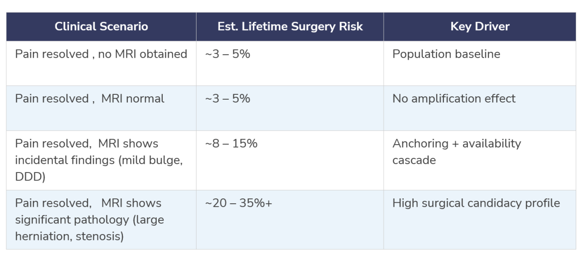
Note: These estimates reflect U.S. population data and published utilization research. Individual risk varies by age, comorbidities, clinical history, and future episode severity. These figures are intended for educational context only and are not individualized medical advice.
The Role of Patient Behavior: How “Finding Something” Changes Everything
It is not only physician behavior that shifts after a documented MRI finding. Patients respond differently too, and that shift in behavior contributes to the elevated surgical risk.
When patients learn they have a disc herniation, stenosis, or degenerative changes on their MRI, even if those findings were never causing their symptoms, several predictable changes tend to occur:
- Patients become more cautious about physical activity, sometimes to a degree that impairs recovery and functional conditioning.
- Fear-avoidance beliefs increase. Patients associate back pain with structural damage rather than with the normal variability of musculoskeletal experience.
- Pain catastrophizing, the tendency to interpret pain signals as more threatening than they are, increases when patients have a structural “explanation” for their discomfort.
- Healthcare utilization rises. Patients with documented findings seek care more frequently, generate more referrals, and are more receptive to escalating interventions.
- Patients begin to perceive themselves as candidates for surgery, sometimes before any clinician has raised the option.
None of this is irrational from the patient’s perspective. If your MRI shows “a herniated disc at L4-5,” it is entirely understandable to interpret that as meaning your spine is damaged. The problem is that this interpretation is frequently incorrect, and the medical system does not always do enough to correct it.
When Spine MRI Is Absolutely the Right Choice
None of the above should be interpreted as discouraging spine MRI when it is clinically indicated. There are clear, well-established situations in which early or urgent MRI is the correct course of action:
- Progressive neurological deficit, worsening weakness, numbness, or coordination difficulties in the arms or legs
- Bowel or bladder dysfunction suggesting cauda equina syndrome, a surgical emergency requiring immediate imaging
- Signs of myelopathy, spinal cord compression producing gait changes, hand clumsiness, or hyperreflexia
- Red flag findings suggesting infection, tumor, or fracture, including fever, unexplained weight loss, trauma history, or cancer history
- Pain that is severe, unrelenting, or unresponsive to six weeks of appropriate conservative care
- Clinical findings that do not fit a musculoskeletal pattern and require further investigation
In all of these situations, the clinical value of MRI is clear, and the benefits of early imaging far outweigh any concerns about downstream anchoring effects. The question this article raises applies to the much more common scenario: acute, non-specific back or neck pain without neurological findings, in a patient who is likely to recover without intervention.
What This Means for You as a Patient
If you are currently recovering from a spine pain episode and your physician has recommended watching and waiting before imaging, that recommendation may be protecting you from more than you realize.
If you have already had an MRI that identified structural findings, that does not mean surgery is inevitable or even likely. It does mean you should understand the following:
- Structural findings on MRI are extremely common in adults and frequently represent normal aging, not injury or disease.
- The presence of a finding does not confirm it is the source of your symptoms.
- Future episodes of back pain are common in the general population regardless of imaging status, and most resolve without surgery.
- Staying active, maintaining core strength, and avoiding catastrophizing your MRI report are among the most evidence-based strategies for long-term spine health.
- Any decision about surgical intervention should be based on a complete clinical picture, a physical examination, functional limitations, and symptom trajectory, not on imaging findings in isolation.

A Final Note from Dr. Shim
In spine surgery, the most important decision is not how to perform an operation, it is whether to perform one at all. The goal of this article is not to alarm patients who have had spine MRIs, nor to suggest that imaging is inherently harmful. It is to ensure that patients and their families have a complete picture of how early imaging decisions can quietly reshape the clinical pathway that follows.
Your spine pain resolved. That is the best possible outcome. The question of what comes next, and whether a prior MRI changes that trajectory, is worth understanding clearly.
If you have questions about your imaging findings, your current symptoms, or whether a surgical consultation is genuinely appropriate in your situation, seek an evaluation with a spine specialist with a reputation for objective analysis without bias toward intervention.
Last modified: June 15, 2026